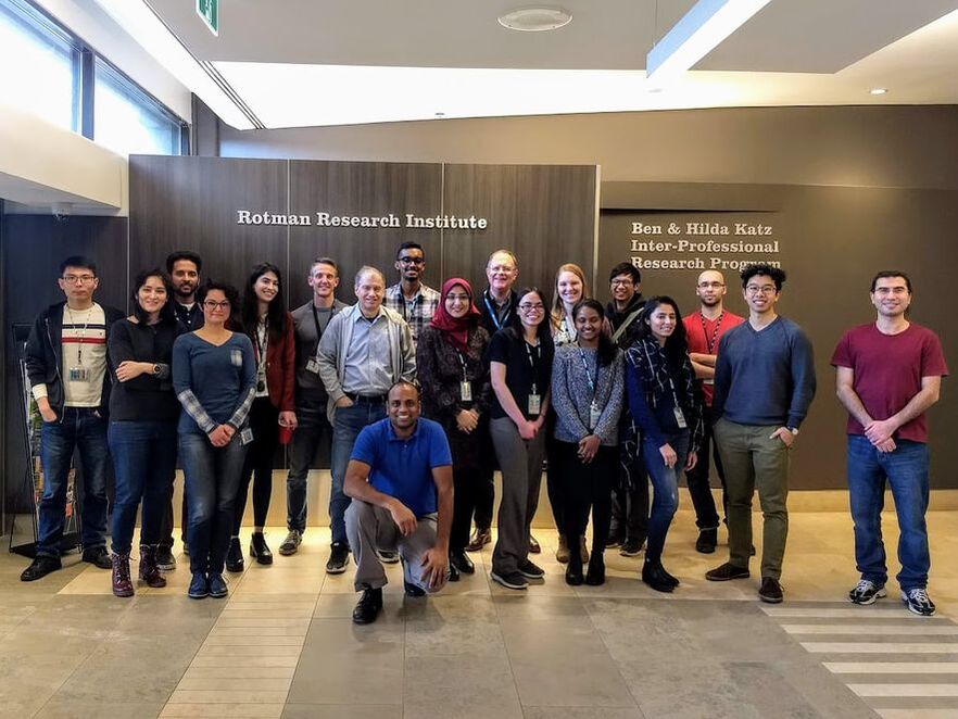
Welcome to Dr. Stephen Strother's lab, located in the Rotman Research Institute at Baycrest Health Sciences Centre. This lab focuses on analysis tools for neuroimaging researchers that are coupled with state-of-the-art neuroimaging research databases. The overall goal is to develop a deeper understanding on human mental functions, and how regular aging, as well as disease can impact the brain.
The Strother Lab is supported by the Baycrest Foundation , the Ontario Brain Institute (OBI), and the multi-institutional stroke program of the Canadian Partnership for Stroke Recovery. To learn more about Dr. Strother, our research, and the members that make up our lab, please check out the links below.
The Strother Lab is supported by the Baycrest Foundation , the Ontario Brain Institute (OBI), and the multi-institutional stroke program of the Canadian Partnership for Stroke Recovery. To learn more about Dr. Strother, our research, and the members that make up our lab, please check out the links below.
OUR FOCUS
Neuroinformatics: is an interdisciplinary research field that focuses on the organization of neuroscience data. This data is usually collected from neuroimaging techniques such as MRI, fMRI, EEG, PET, MEG and CT scans, and then organized and analyzed using computational models and research analytical tools. Neuroinformatics also involves finding suitable ways to store and share large datasets for collaboration and analysis. In the Strother lab we manage a neuroimaging database named SPReD for this very purpose. For more information on our neuroinformatics projects please click here.
Functional MRI Preprocessing and Analysis Optimization: involves correcting for distortion in brain images caused by artifacts such as; metal (often from dental implants), physiological noise, and participant motion while under the fMRI. With the use of preprocessing we can help optimize signal activation and become more certain that the signal found is indeed due to brain activation and not just due to artifact. In the Strother lab we create and use preprocessing pipelines to help clean up data. For more information on this please click here.
Multimodal Imaging: involves combining two or more imaging modalities, usually within a single imaging session. This technique may provide an improved solution to overcoming the limitations of an independent modality, while expanding the information provided. One example of this in our lab includes simultaneously recorded EEG-fMRI data.
|
Rotman Research Institute | Baycrest
3560 Bathurst Street, North York Ontario, Canada M6A 2E1 |


